
Oblique Cross-Section of the Eye: Case Study
Oblique Cross-Section of the Eye: Case Study
Media
Cinema 4D, ArtStudio Pro, Adobe Illustrator
Target Audience
Anatomy students
Client
Michael Corrin
Context
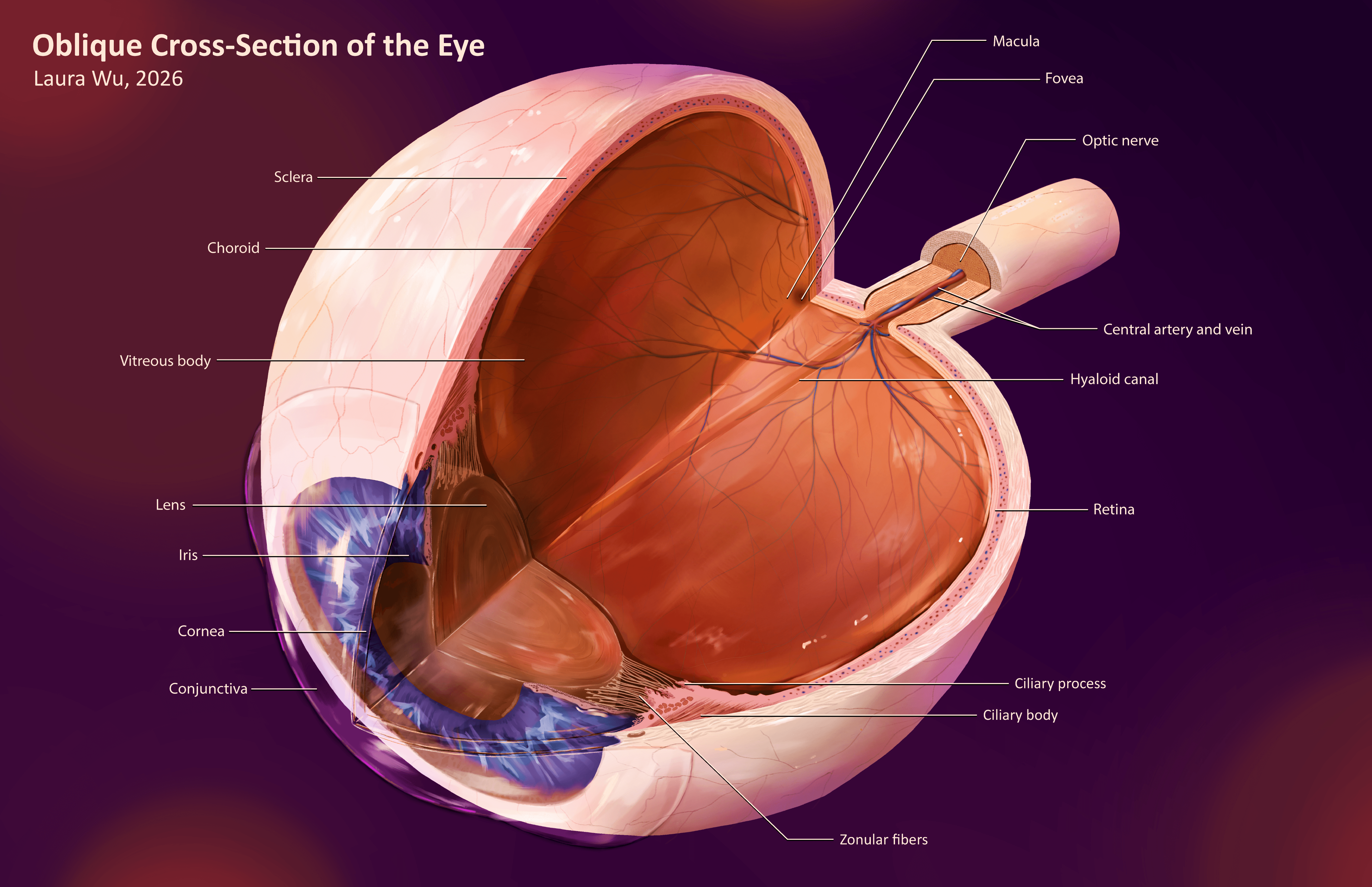
For this project, my teammates and I created individual illustrations of key internal structures of a human eye through a cross-sectional view.
Process
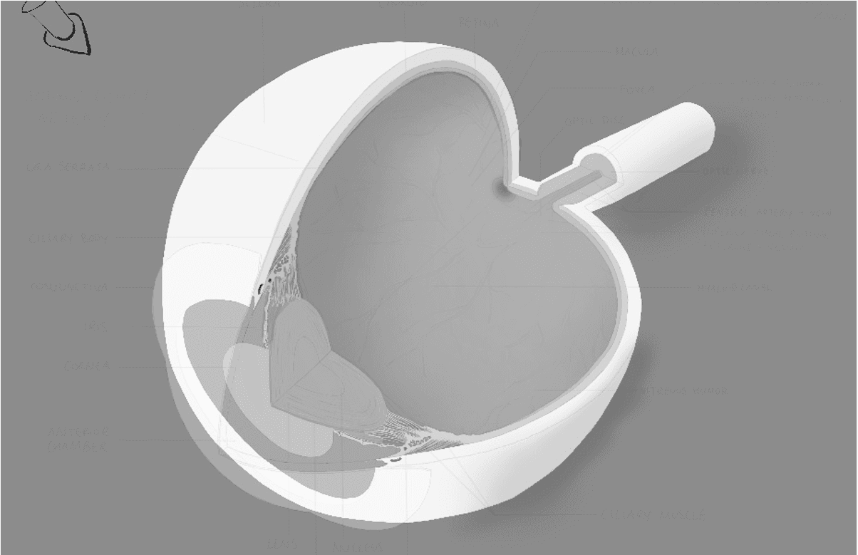
At the outset of the project, our team began by collecting references from atlases, textbooks, journal articles, and websites that provided detailed information about the internal and external structures of the eye. A complete list of references can be found at the end of this document. The purpose of this preliminary research was to prepare us to build our own accurate 3D models of the eye, which we would be able to freely manipulate prior to creating our 2D illustrations. Our approach to the maquettes involved a 2D-to-3D workflow: we created splines in Adobe Illustrator and imported them into Cinema4D to be lathed and staged. My initial maquette featured staggered layers of the right eye viewed from a superior-oblique angle.
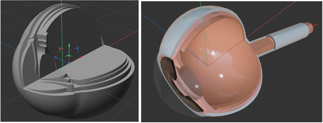
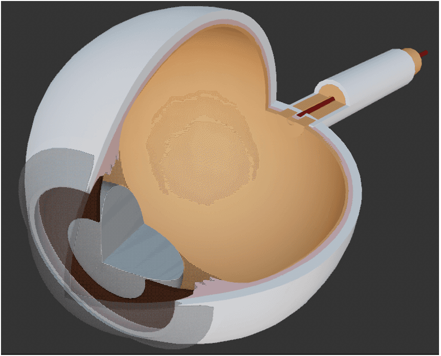
After reviewing each other’s work and receiving feedback, I revised my maquette to remove the staggered layers and position the eye from a more anterior view. The rationale for this decision was to reduce the complexity of the illustration, i.e. the amount of texture that each individual layer would require to be rendered and the strategic avoidance of rendering the ora serrata. I also revised the accuracy of my maquette by double-checking the quantitative measurements in my spline and adding the conjunctiva to the model. I produced a second draft that would become the basis for my illustration. To be efficient with my time, I decided to only include the structural foundations of the eye in my maquette. I judged this to be sufficient for beginning the illustration as I would render the details, texture, and lighting by hand later on.
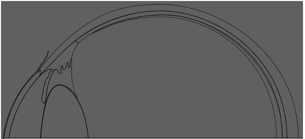
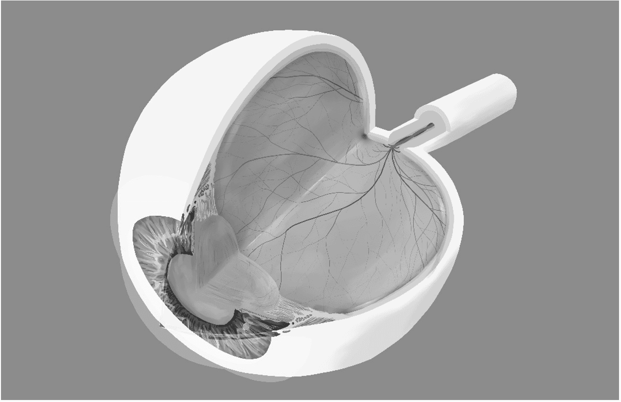
The program that I used to illustrate this project was ArtStudio Pro. I developed several rough drafts of my sketch by tracing over the maquette and labelling the core elements of the eye. Iterative feedback helped me refine the rough draft into the final version that would be used as the basis for rendering the illustration:
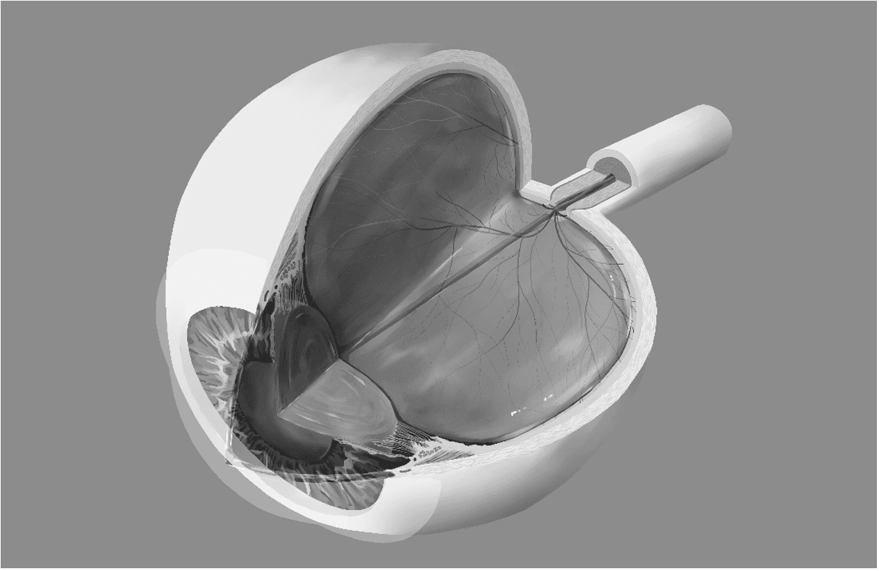
Once the linework for the sketch was finalized with as much detail as possible, I began the process of rendering it in grayscale by blocking in tentative values for each layer. I established an upper lefthand light source that was slightly angled towards the front of the eye, and I chose a darker background for this piece to increase contrast and visual salience.
Texturing individual surfaces of the eye primarily required references from real photos or micrographs (light microscopy or electron microscopy) where available, which I supplemented with clear diagrams. I worked from the anterior to the posterior chambers.
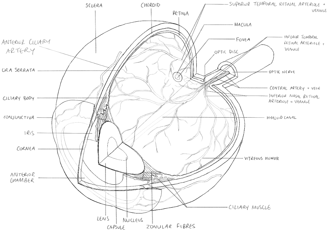
I first rendered the cross-sections of the iris, ciliary body, and lens. Referring to my collected resources, I included the sphincter/dilator pupillae muscles, pigment layer, minor/major arterial circles, iridial folds, pupillary margin, and trabecular meshwork of the iris; the circular and longitudinal ciliary muscles in the ciliary body; the canal of Schlemm in the sclera; and the wispy texture of the ciliary processes and the depth of the zonular fibers in between them. I then blocked out the path of the arterioles and venules in solid color, verifying the proportional thicknesses of the branches relative to the central vessels as documented in the literature. Furthermore, I examined the reflective and glossy properties of the lens and vitreous humor from the reference images (in the absence of a real animal eyeball) and mapped out the values for the lighting that I pictured for this piece.
Once I completed the grayscale render, I colorized the illustration using different blending modes. I chose an analogous color scheme for the entire piece.
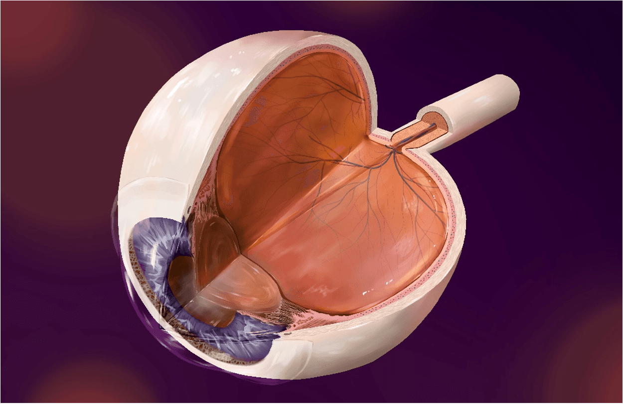
After receiving feedback, I clarified the boundaries of the anterior chamber, cornea, and conjunctiva with clearer values and lines. I also decreased the opacity of the trabecular meshwork after referring to photos of our own eyes and realizing that the transition in the limbus was soft instead of crisp. Once the image was finalized, I imported it into Adobe Illustrator to label the significant components of the eye.
Media
Cinema 4D, ArtStudio Pro, Adobe Illustrator
Target Audience
Anatomy students
Client
Michael Corrin
Context
For this project, my teammates and I created individual illustrations of key internal structures of a human eye through a cross-sectional view.
Process
At the outset of the project, our team began by collecting references from atlases, textbooks, journal articles, and websites that provided detailed information about the internal and external structures of the eye. A complete list of references can be found at the end of this document. The purpose of this preliminary research was to prepare us to build our own accurate 3D models of the eye, which we would be able to freely manipulate prior to creating our 2D illustrations. Our approach to the maquettes involved a 2D-to-3D workflow: we created splines in Adobe Illustrator and imported them into Cinema4D to be lathed and staged. My initial maquette featured staggered layers of the right eye viewed from a superior-oblique angle.
After reviewing each other’s work and receiving feedback, I revised my maquette to remove the staggered layers and position the eye from a more anterior view. The rationale for this decision was to reduce the complexity of the illustration, i.e. the amount of texture that each individual layer would require to be rendered and the strategic avoidance of rendering the ora serrata. I also revised the accuracy of my maquette by double-checking the quantitative measurements in my spline and adding the conjunctiva to the model. I produced a second draft that would become the basis for my illustration. To be efficient with my time, I decided to only include the structural foundations of the eye in my maquette. I judged this to be sufficient for beginning the illustration as I would render the details, texture, and lighting by hand later on.
The program that I used to illustrate this project was ArtStudio Pro. I developed several rough drafts of my sketch by tracing over the maquette and labelling the core elements of the eye. Iterative feedback helped me refine the rough draft into the final version that would be used as the basis for rendering the illustration:
Once the linework for the sketch was finalized with as much detail as possible, I began the process of rendering it in grayscale by blocking in tentative values for each layer. I established an upper lefthand light source that was slightly angled towards the front of the eye, and I chose a darker background for this piece to increase contrast and visual salience.
Texturing individual surfaces of the eye primarily required references from real photos or micrographs (light microscopy or electron microscopy) where available, which I supplemented with clear diagrams. I worked from the anterior to the posterior chambers.
I first rendered the cross-sections of the iris, ciliary body, and lens. Referring to my collected resources, I included the sphincter/dilator pupillae muscles, pigment layer, minor/major arterial circles, iridial folds, pupillary margin, and trabecular meshwork of the iris; the circular and longitudinal ciliary muscles in the ciliary body; the canal of Schlemm in the sclera; and the wispy texture of the ciliary processes and the depth of the zonular fibers in between them. I then blocked out the path of the arterioles and venules in solid color, verifying the proportional thicknesses of the branches relative to the central vessels as documented in the literature. Furthermore, I examined the reflective and glossy properties of the lens and vitreous humor from the reference images (in the absence of a real animal eyeball) and mapped out the values for the lighting that I pictured for this piece.
Once I completed the grayscale render, I colorized the illustration using different blending modes. I chose an analogous color scheme for the entire piece.
After receiving feedback, I clarified the boundaries of the anterior chamber, cornea, and conjunctiva with clearer values and lines. I also decreased the opacity of the trabecular meshwork after referring to photos of our own eyes and realizing that the transition in the limbus was soft instead of crisp. Once the image was finalized, I imported it into Adobe Illustrator to label the significant components of the eye.